Segond fracture constitute a special challenge for both physical therapists (PTs) and occupational therapists (OTs) in the ever-changing field of healthcare. When it comes to the healing and rehabilitation of patients with Segond fractures and related knee injuries, these specialists are indispensable. We will delve into the realm of Segond fractures in this extensive post, covering their etiology, symptoms, diagnosis, surgical intervention, and above all the critical role that physical therapists and occupational therapists play in the healing process.
Understanding the Segond Fracture
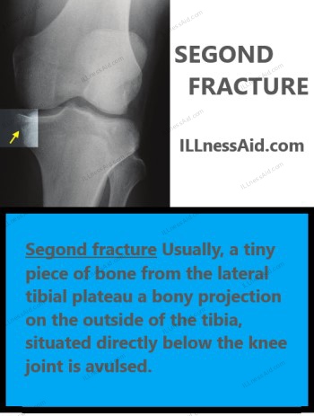
A particular kind of knee injury known as a “Segond fracture,” named for the French surgeon Paul Segond, frequently happens in conjunction with anterior cruciate ligament (ACL) rupture. Usually, a tiny piece of bone from the lateral tibial plateau a bony projection on the outside of the tibia, situated directly below the knee joint is avulsed in these fractures. Segond fractures typically occur as a result of an abrupt, twisting action of the knee joint along with an extension force.
These fractures are noteworthy due to their association with ACL damage in addition to their clinical significance. Damage to the ACL, a crucial ligament that stabilizes the knee joint, can result in significant functional deficits. As a result, PTs and OTs must comprehend Segond fractures and how to treat them.
Causes and Mechanism of Segond Fractures
Segond fractures are frequently brought on by the ACL experiencing too much stress and strain when engaging in activities that require abrupt direction changes or pivoting. The lateral tibial plateau fragment may avulse from the bone as a result of this tension. Sports-related injuries, like those sustained in football, soccer, and skiing, where players routinely make sudden pauses and direction changes, are common causes of Segond fractures. This kind of injury can cause simultaneous ACL damage and a Segond fracture.
Symptoms of Segond Fractures
A variety of symptoms can greatly affect a patient’s day-to-day functioning after suffering a Segond fracture. These signs and symptoms are frequently strongly linked to the ACL tear that accompanies the fracture. Typical indications and symptoms include of:
- Knee Pain: Patients often complain of acute, excruciating pain in the vicinity of their knees, especially on the lateral side.
- Swelling: Swelling, also known as edema, is a typical reaction to injuries to the knee and can appear hours after the injury.
- Limited Range of Motion: Patients may find it difficult to move their knee and may notice a reduced range of motion as a result of pain and swelling.
- Instability: One common symptom of ACL involvement is an unstable knee that gives way when walking or bearing weight.
- Popping or Snapping Sensation: A lot of patients report feeling or hearing a pop or snap at the scene, which could be a sign of tendon or ligament damage.
Diagnosing Segond Fractures
To guarantee the right course of treatment, a precise diagnosis of Segond fractures is essential. A patient’s medical history, physical examination, and diagnostic imaging are usually used in conjunction to make a diagnosis. Healthcare professionals will measure the patient’s range of motion, and joint stability, and conduct particular tests to determine the ACL’s integrity during the physical examination. The pivot-shift test, the anterior drawer test, and the Lachman test are a few examples of these tests.
1. Lachman Test
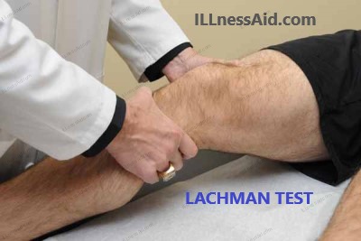
- Goal: One of the main ligaments in the knee joint, the ACL, is evaluated for integrity using the Lachman test. It aids in the diagnosis of ACL damage or tears.
- Procedure: The patient usually lies on their back, or in a supine position. Using one hand to stabilize the femur, the examiner holds the patient’s thigh, and with the other, they grasp the patient’s lower leg, or tibia, just below the knee joint. The examiner then feels for any abnormal movement while attempting to move the tibia forward in relation to the femur.
- Interpretation: An ACL injury is likely present in a positive Lachman test result. Examiners typically detect abnormally increased forward movement of the tibia when there is an ACL tear. The patient may experience pain or discomfort during the test.
2. Anterior Drawer Test
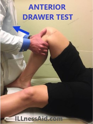
- Goal: The anterior drawer test evaluates the stability of the ACL, much like the Lachman test does.
- Procedure: The patient usually lies on his or her back with the knee bent 90 degrees. To support the patient’s lower leg, the examiner perches on their foot. The examiner tries to pull the tibia forward by grasping the patient’s lower leg just below the knee joint with both hands.
- Interpretation: Similar to the Lachman test, a positive anterior drawer test indicates an ACL injury. ACL instability is indicated by excessive forward movement of the tibia combined with pain or discomfort.
3. Pivot-Shift Test
- Goal: The pivot-shift test is intended to identify ACL injuries, but it is also helpful in evaluating functional instability and the possibility of abnormal joint movement, or knee subluxation, during dynamic activities.
- Procedure: The examiner flexes the patient’s hip and knee to a range of 20 to 30 degrees while the patient is usually in a supine position. Next, the examiner places one hand on the patient’s heel and the other on the patient’s tibia. The examiner rotates the knee and extends the tibia while applying a valgus (inward) force. This set of movements mimics the action of a pivot.
- Interpretation: When the pivot-shift test is positive, the tibia moves abnormally during the maneuver, causing a characteristic “shift” or “clunk” sensation. It can be especially relevant for athletes or people whose activities require a high level of knee stability because it shows both potential functional instability and ACL deficiency.
Diagnostic imaging is an essential part of the process. X-rays can be used to assess the alignment of the knee’s bones and spot fractures. However, Segond fractures might not always show up on X-rays, necessitating additional imaging tests like computed tomography (CT) scans or magnetic resonance imaging (MRI). ACL tears and other injuries can be detected more easily thanks to these sophisticated imaging techniques, which offer a more detailed view of soft tissues and ligaments.
Treatment for Segond fracture
Role of PT in Segond fracture
When it comes to the rehabilitation of patients who have had surgery for Segond fractures and the resulting ACL damage, physical therapists are essential. Their knowledge of movement and functional rehabilitation is extremely helpful in assisting patients in regaining strength and mobility.
To promote a safe and efficient recovery, physical therapists (PTs) work with patients right after surgery. Usually, the goals of this stage are to control pain, minimize swelling, and avoid side effects like blood clots. Exercises involving a passive range of motion can be started to avoid stiffness.
Physical therapists lead patients through a series of exercises to increase strength, stability, and range of motion while the knee heals. The effectiveness of ACL reconstruction depends on these exercises. In order to help patients regain an awareness of joint positioning, therapists may also focus on proprioception, balance, and neuromuscular training.
PTs concentrate on assisting patients in regaining functional strength and movement in the later phases of rehabilitation. To guarantee a safe return to their activities, athletes frequently receive training tailored to their particular sport.
PTs are essential in teaching patients self-management techniques and ways to avoid getting hurt again during these phases. This instruction includes advice on how to maintain appropriate body mechanics, apply strengthening exercises, maintain a healthy lifestyle, and use ergonomics. The goal is to provide patients with the tools like kneecaps they need to lower their risk of developing new knee injuries and to enhance their long-term well-being.
Role of OT in Segond fracture
Occupational therapists (OTs) are crucial in assisting patients in regaining their independence and functionality in their daily lives, while physical therapists (PTs) focus primarily on the physical aspects of recovery. This is particularly important for people who have had surgery for Segond fractures because their knee injury may make it difficult for them to perform daily tasks. OTs responsibilities include:
- ADL TRAINING: Occupational therapists (OTs) work with patients to ensure they are able to independently perform activities of daily living (ADLs), such as dressing, grooming, and bathing.
- Assistive Device Training: Occupational therapists (OTs) offer instruction and direction on the safe and efficient use of mobility aids such as crutches, canes, and walkers to patients who need them.
- Home Modifications: Occupational therapists evaluate their patients’ living spaces and suggest adjustments as needed to meet their physical needs.
- Cognitive and Psychological Support: Patients may occasionally suffer from depression or anxiety as a result of their injury. OTs can offer techniques and assistance to deal with these emotional difficulties.
- Return to Work Planning: OTs help create a plan that takes into account the physical capabilities and unique job requirements of individuals who have suffered Segond fractures and must return to work.
Exercises For Segond Fracture
Physical therapists (PTs) and Occupational therapists (OTs) usually recommend these exercises to enhance knee joint strength, stability, and mobility. To guarantee correct form and safety, patients should carry out these exercises under the supervision of a medical professional. Please be aware that the particular exercises and how they progress may change based on the condition and treatment plan of the individual:
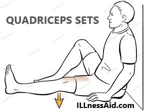
- Goal: Building strength in the quadriceps muscle group aids in knee stabilization.
- How to do it: Tighten the muscles in your thighs while sitting or lying down, pressing the back of your knee toward the floor. After a brief period of holding, release. Continue for multiple sets.
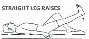
- Goal: This exercise strengthens the quadriceps and enhances knee stability.
- How to do it: Raise the straight leg a few inches off the ground while maintaining its straight position while lying on your back with the other leg bent. After holding it for a short while, lower it. Make sure to repeat this exercise several times.
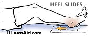
- Goal: Promotes range of motion and flexibility in the knee.
- How to do it: While on your back, bend your knee and slide your heel toward your buttocks. Return your heel to the starting position with gentle pressure. This motion should be repeated several times.
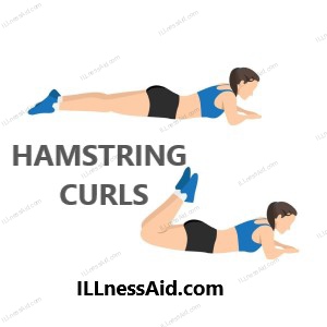
- Goal: Builds the hamstring muscles at the rear of the thigh, which aids in knee support and balance.
- How to do it: Curl your legs in the direction of your buttocks or by using a leg curl machine or resistance bands. Repeat for several sets after letting go.
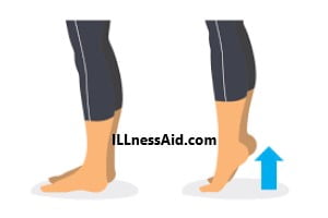
- Goal: Improves calf stability and strength, which helps support the knee joint.
- How to do it: Place your heels hanging over the edge of a step or a sturdy platform. Lift your heels below the step and then push up onto your toes. Do this several times over.

Conclusion
In summary, the holistic approach taken by physical therapists (PTs) and occupational therapists (OTs) is critical to the recovery process of patients with Segond fractures and related ACL injuries. Patients can overcome difficult obstacles, regain their strength and mobility, and eventually look forward to a future filled with improved health and well-being with the help of these professionals’ expertise. By addressing the physical, functional, and emotional aspects of recovery, the patient-centered and collaborative approach enables individuals to lead fulfilling lives after their injury.
Thank you for reading this far, today we talked about segond fracture and its PT & OT management with exercises. If you have any doubts regarding this then Contact us.